Rhegmatogenous Retinal Detachment Assignment Sample
The following assignment sample work shall highlight the condition of (RRD) Rhegmatogenous Retinal Detachment, symptoms of the retinal detachment, and diagnosis and treatments of RRD condition.
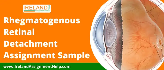
Retinal detachment is described as an emergency situation in which the retina tissue at the back of the eye gets pulled away from its normal position. A layer of blood vessels responsible for providing nourishment and oxygen for the eyes gets separated from retinal cells. Such displacement of the retina can even cause a permanent loss of vision.
There are mainly three causes of retina detachment and they are tractional, exudative, and rhegmatogenous. This is a very common condition among people who have a family history of retina detachment, are middle-aged, have previous eye surgery, or have any severe eye disease and injury.
In this assignment sample, we will discuss the Rhegmatogenous Retinal Detachment in detail.
Condition of Rhegmatogenous Retinal Detachment
Rhegmatogenous is a common type of retinal detachment which is caused due to a tear in the retina that allows the fluid to pass through and get collected under the retina which pulls away from the retina from the underlying tissue. When the blood supply gets restricted into the area of the detached retina it can lead to a situation of permanent loss of vision.
The most common cause of this condition is considered to be age. It is the separation of the neurosensory retina from retinal pigment epithelium through fluid transversing from the vitreous cavity into subretinal space.
Hire an Irish Assignment Writer to Write your Essay, Thesis & Other Academic Papers
Symptoms of Retinal Detachment
There are many definite signs of retinal detachment in a person but are clearly only visible when such a condition gets into an advanced stage. The common symptoms or signs of retinal detachment are:-
- Flashes of light in one or both the eyes.
- Blurred vision.
- Shadowed vision in the eyes.
- The sudden appearance of floaters and tiny specks drifting the field of vision.
- Gradually reduced side (peripheral) vision.
Diagnose and Treatments of Rhegmatogenous Retinal Detachment
Since the detachment is unpredictable and can vary its timings but if tears are caught early then it can be treated with a laser in a clinical setting. If once the rhegmatogenous retinal detachment is developed surgical intervention gets necessary.
Symptomatology
When a patient visits an eye care specialist the retinal detachment gets easily detected by him. An asymptomatic retinal detachment is a temporal detachment arising from the tears.
In the symptomatology process, there is a collection of symptoms that is closely studied by the specialist to diagnose the issue. There are many causes of RRD where the patients is not able to notice flashes if they are asleep so the unilateral loss of vision is ignored and detachment is noticed on getting a blurred central vision.
Floaters are common symptoms of this condition for all age groups. There will be complaints of flashes, floaters, or negative dyschromatopsia where these symptoms are associated with the RRD.
Negative dyschromatopsia may cause flashes and loss of vision and a decrease in peripheral and central vision. After the study of such collective symptoms, the clinician can refer the patient to an eye specialist.
Scared with Looming Deadline, Buy Plagiarism Free Paper Writing Services Now
Clinical Evaluation
The other way of detecting the condition of rhegmatogenous retinal detachment can be done with the help of clinical evaluation. A macula involving retina detachment is easy to identify and spot but a peripheral detachment cannot be easily detected.
Any clue of subretinal fluid or retinal break can be an immediate sign of reference to a doctor before visiting any such specialist.
If the clinical evaluation does not come out to be successful in the detection of retinal detachment then three major contact lens examinations and scleral depression can help in the detection of peripheral tears. Vitreous hemorrhage or pigment cells in the vitreous can increase the risk of retina detachment.
In such cases, ultrasonography or vitrectomy is required and any careful examination can detect PVD or vitreous hemorrhage causing the break in the retina. There might be other conditions as well that may arise such as neoplastic disease, tractional retina detachment, and inflammation.
Auxiliary Testing
Auxiliary testing is considered to be an effective technique to diagnose the RRD condition with the help of dilated fundus exam. These tests help in identifying the severity of retinal abnormalities. Widefield photography can help with scanning RRD.
Optical Coherence Tomography (OCT) in the widefield mode is very helpful in the diagnosis of RRD and can assess the macular status at the time of diagnosis. Using B-scan ultrasound is recommended only when there is no clear clinical view must be done in presence of media opacification.
In a few cases, Fluorescein Angiography and Visual Field Testing can also be used the diagnose RRD.
Get 100% Unique Assignment Papers for Your College & Get Good Grades
Laser Treatments and Pneumatic Retinopexy
Laser treatments are effective only in those regions where the neurosensory retina and RPE can be brought together through scleral depression. A laser is applied to the border of the detached retina.
All other retinal defects can be treated through laser. Prophylactic retinopexy is a low-risk treatment for a person with this condition.
Pneumatic retinopexy can be used as the best treatment of RD where the retina break is located if more than one bubble exists they are localized in such a way so that the expansive bubble can adequately cover them with the proper position of the patient.
Retinopexy can be performed by cryotherapy by applying a laser to the breaks in prior to gas injection which is performed after several hours or days once the retina gets attached.
Online Writing Guidance at the Most Convenient Price for the Irish College Students!
The above-written assignment sample is based on the healthcare-related topic of Rhegmatogenous Retinal Detachment.
Healthcare QQI Level 5, Skincare and Eye Treatment QQI Level 5, Nursing QQI Level 5, Medical Terminology and Administration Course QQI Level 5, Wellbeing QQI Level 5students can read this assignment sample to gain knowledge on such healthcare topics.
We have many healthcare, disease, and medical essay samples available on our official website of Ireland assignment help which can be read by the students for the purpose of reference.
Our online assignment writing services are available for you 24×7 at just a call away. Experts of our Irish writing company can provide their writing services at the most convenient price for you.
Hire an Irish Assignment Writer to Write your Essay, Thesis & Other Academic Papers
- 5N1794 Safety and Health at Work Assignment Sample Ireland
- 5N0758 Care Support Assignment Sample Ireland
- 5M4339 Healthcare Support Assignment Sample Ireland
- 5M4468 Community and Health Services Assignment Sample Ireland
- GP5101 Cardiovascular Disease in Primary Care UCC Assignment Sample Ireland
- Cognitive Behavioural Modalities Assignment Sample Ireland
- Psychodynamic Modalities Assignment Sample Ireland
- Integrative Modalities Assignment Sample Ireland
- SP139 Community Health Assignment Sample NUIG Ireland
- STN1107 Professional Practice Assignment Sample NUI Galway Ireland
- STN1106 Primary and Community Mental Health Care Assignment Sample NUI Galway Ireland
- AN230 Human Body Structure Assignment Sample NUIG Ireland
- CR6801: Trauma and Victimology Assignment Sample Ireland
- Equipment and Caring Aids that are part of the Irish Healthcare Essay Example Ireland
- Down Syndrome Essay Example Ireland
